Fellow leads research that could catapult mechanobiology advances into the clinical arena
Engineering Fellow Dr Thierry Savin led a research team that has developed a new electromagnetic device that enables high-precision measurements of a broad range of soft biological tissues. The team say that it has established a new standard of precision in the mechanobiology field.
The method allows for the mechanical testing of tissues the size of human biopsy samples, making it particularly relevant for studies of human disease.
The body’s soft tissues exhibit a wide range of mechanical properties, such as stiffness and strength, which are critical to carry out their function. For example, the tissues of the gastrointestinal tract are soft to allow for the transit and digestion of food, whereas tendons are relatively more stiff to transmit force from muscle to bone, allowing us to move.
The ability to accurately measure the mechanical properties of these tissues, which are subject to change during developmental processes or because of disease, has profound implications for the fields of biology and medicine. Methods to measure these properties are currently inadequate, and their accuracy and reliability remains limited – until now.
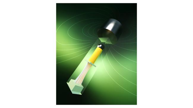
New research involving researchers from the University of Cambridge and the MIT Institute for Medical Engineering and Science (IMES) has resulted in a device that relies on magnetic actuation and optical sensing, thus potentially allowing for live imaging of the tissue under an inverted microscope. This way, insights can be gained into the behaviour of the tissue under mechanical forces at both a cellular and molecular level. The results are reported in the journal Science Advances.
An electromagnet exerts a pulling force on the tissue specimen which is mounted on the device, while an optical system measures the specimen’s change in size or shape.
“One of the most critical requirements for mechanical testing of soft biological tissues is the need to mimic the biological specimen’s physiological conditions (e.g. temperature, nutrients) as closely as possible, in order to keep the tissue alive and preserve its biomechanical properties,” said Thierry, who is also a University Associate Professor in Bioengineering. “To this end, we designed a transparent mounting chamber to measure the mechanical properties of tissues – at the millimetre scale – in their native physiologic and chemical environment. The result is a more versatile, precise, and robust device that shows high reliability and reproducibility."
To directly assess the performance of their electromagnetic device, the researchers conducted a study on the biomechanics of a mouse oesophagus and of its constitutive layers. The oesophagus is the muscular tube connecting the throat with the stomach and it is composed of multiple tissue layers. The researchers used the device to conduct the first biomechanical investigation of each of the three individual layers of the mouse oesophageal tissue. Their findings showed that the oesophagus behaves like a three-layer composite material akin to those commonly used in several engineering applications. To the researchers’ knowledge, these are the first results acquired of the mechanical properties of each individual layer of the oesophagus.
“Our study demonstrated the enhanced reliability of the electromagnetic device, yielding errors in the stress-strain response below 15% – a level of accuracy not seen before,” said Dr Adrien Hallou, Postdoctoral Fellow at the Wellcome Trust/Cancer Research UK Gurdon Institute. “We hope that this device may eventually become the new standard in the tissue biomechanics field, providing a standardised dataset for the characterisation of mouse and human soft tissue mechanics across the board.”
Reference:
Luca Rosalia; Adrien Hallou; Laurence Cochrane; Thierry Savin. ‘A magnetically actuated, optically sensed tensile testing method for mechanical characterization of soft biological tissues’. Science Advances (2023). DOI: 10.1126/sciadv.ade2522
This is based on an article originally published by the Department of Engineering. It is reproduced under a Creative Commons Attribution 4.0 International License.